Sweet Poison, Star fruit neurotoxin identified, caramboxin

 Star fruit neurotoxin identified
Neurotoxin from Star Fruit
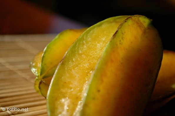
Patients with kidney disease have to watch what they eat: bananas, oranges, tomatoes, nuts, broccoli, and beans are all off-limits. Putting star fruit or carambola on the menu would be downright dangerous. This fruit contains a substance that is a deadly neurotoxin for people with kidney disease. Brazilian researchers have now isolated and identified this neurotoxin. As they report in the journal Angewandte Chemie, it is an amino acid similar to phenylalanine.
Caramboxin: Patients suffering from chronic kidney disease are frequently intoxicated after ingesting star fruit. The main symptoms of this intoxication are named in the picture. Bioguided chemical procedures resulted in the discovery of caramboxin, which is a new phenylalanine-like molecule that is responsible for intoxication. Functional experiments in vivo and in vitro point towards the glutamatergic ionotropic molecular actions of caramboxin, which explains its convulsant and neurodegenerative properties.
Sweet Poison
star fruit (Averrhoa carambola) or carambola has been cultivated in Malaysia, Southern China, Taiwan, India and Brazil. It is rather popular in the Philippines and Queensland, Australia and moderately so in some of the South Pacific Islands, particularly Tahiti, New Caledonia and Netherlands New Guinea, Guam and in Hawaii and south Florida. There are some subspecies in the Caribbean Islands, in Central America and in tropical West Africa. The fruits are also available in many European countries and Canada. Range of soluble oxalate salts concentrations obtained from many cultivars varies from 80 to 730 mg/100 g of the fruit
Structural Elucidation and Spectroscopic Data of Caramboxin (1)
Most 1H and 13C NMR data from the isolated compound were easily assigned due to the relationship
of this compound with well-known aromatic amino acids such as phenylalanine. The side chain is
identical to phenylalanine as attributed in the NMR data below. The tetra-substituted pattern of the aromatic ring was also easily recognized by the only two signals for aromatic protons at δ 6.42 and6.37 with a coupling constant (2.0 Hz) typical of aromatic meta coupling. The positioning of
substituents in the aromatic ring were attributed due to the 13C chemical shifts and confirmed by
HMBC experiment. In this way acetyl group was placed at C-6 due to the low chemical shift (104.1ppm) of this aromatic carbon; methoxyl was placed at C-3 due to its highest chemical shift (165.2ppm) among aromatic protons, which was confirmed by HMBC experiment; finally the hydroxyl group was placed at the remaining C-5. Comparison of the observed accurate mass measurement with theoretically calculated formulae for the signal at m/z 256.0823 allows only a few reasonable [MH]+ ion formulae within a standard deviation of 50 ppm. The ion formula [C11H13NO6 + H]+ had the best mass accuracy (0.7 ppm error) and correlation with the NMR spectra. The MS/MS spectrum of m/z 256 shows an intense ion at m/z 192 in addition to neutral elimination of H2O (m/z 238) and CO2 (m/z 212) from the carboxylic acid group. In source dissociation followed by MS/MS analysis revealed that m/z 192 was only obtained from m/z 238, due to the elimination of CH2O2 (46 massunits) as a neutral molecule by 1,2 elimination. The same mechanism was also observed for the m/z166 formation from the ion at m/z 212. Finally, 2D NMR data from HMQC and HMBC confirmed the entire spectral assignment
Caramboxin: 1H-NMR (400 MHz, D2O) δ 6.42 (d, J = 2.0 Hz, 1H, H-4), 6.37 (d, J = 2.0 Hz, 1H, H-2), 4.25 (dd, J = 5.5, 8.0 Hz, 1H, H-8), 3.80 (s, 3H, H-11), 3.66 (dd, J = 14.0, 5.5 Hz, 1H, H-7A),3.18 (dd, J = 14.0, 8.0 Hz, 1H, H-7B);
13C-NMR (100 MHz, DMSO-d6) δ 172.8 (C, C-9), 171.2 (C,C-10), 165.2 (C, C-5), 162.2 (C, C-3), 138.8 (C, C-1), 110.0 (CH, C-2), 104.1 (C, C-6), 100.5 (CH,C-4), 55.4 (CH3, C-11), 53.5 (CH, C-8), 35.9 (CH2, C-7); 15N-NMR (50 MHz, DMSO-d6) δ -268(nitromethane as internal reference).
HMBC (500 MHz, DMSO-d6): H-2 → C-3, C-4, C-7; H-4 →C-2, C-3, C-5; H-7 → C-1, C-2, C-8, C-9; H-8 → C-1, C-7, C-9; H-11 → C-3;
HRMS (m/z): [MH]+calcd for C11H14NO6, 256.0816; found: 256.0818

No comments:
Post a Comment